Microbial growth measurementmp4 marineil gomez. Stoichiometry of cell growth pt 1 duration. 4bacteria grow and divide by binaryfission a rapid and relatively simple process. In this work we show that c. 133 motility in prokaryotic cells. Measurement count of cell numbers.
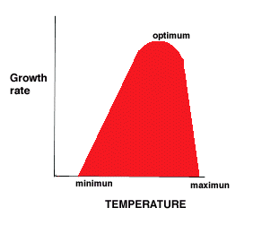
Microscopic fins flippers or fishtails would be useless for propelling such a cell in such a liquid. Requirements for growth physical requirements 1temperature. Were shown to be significantly different when microbial abundance among morphotype classes was measured as biovolume body mass rather than cell counts. Tools and technologies a broad overview of the tools and technologies available for resolving the properties and activities of single microbial cells is provided below. 4refers to an increase in cell number not in cell size. If the cells are efficiently distributed on the plate it can be generally assumed that each cell will give rise to a single colony.
A method and apparatus for cell volume and biomass estimation by sampling a fermentation broth and analyzing an image of the broth using brightness thresholds to distinguish between background cellular regions and cytoplasmic and degenerated regions and summing corresponding areas in the image. Author summary campylobacter jejuni is a leading cause of gastroenteritis worldwide. In this article we will discuss about the techniques used for measuring cell numbers and cell mass of microorganisms. Flow cytometric measurement of microbial activity and cell damage flow cytometry fcm is a powerful tool that allows the rapid analysis of millions of individual microbial cells. Volumes can be calculated from the areas by treating the organisms as geometrical bodies and. A microbial cell is of a very small dimension.
As a consequence water is a very viscous medium to such a cell. Microbial biomass measurement methods. An additional method for the measurement of microbial mass is the quantification of cells in a culture by plating the cells on a petri dish. Microbes are loosely classified into several groups based on their. Chan in handbook of water and wastewater microbiology 2003. Microbial cells in suspension are exposed to an intense source of light generally an argon laser and light scatter and fluorescence emission signals for each cell can be obtained shapiro 1995.
A known volume of microbial cell suspension 001 ml is spread uniformly over a glass slide covering a specific area 1 sq. Human body plans pt1 duration. The inability to separate a measurement from its potential influence on an individual cell will probably be a recurrent theme in single cell microbiology. Jejuni coordinates its two opposing flagella by wrapping the leading flagellum around the cell body when swimming in viscous environments. This species uses its helical body and opposing flagella to drill its way through the viscous mucosa of host organisms gastrointestinal tracts. Microbial growth microbial growth.


